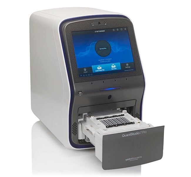

The QuantStudio 7 Pro DNA Amplifier for Quantitative PCR provides reliability, accuracy and sensitivity for your experiments with interchangeable block options: 96-well 0.1 or 0.2 ml, 384-well and TaqMan Array cards.
Blocks are replaced by simply pressing a button or using a voice command. The system comes with one block type, other block types can be purchased separately. Orbitor Microplate Mover (sold separately) is included to support high-performance workflows.
Each tool is equipped with face authentication login, microphone for voice commands, speakers for voice command feedback and instructional videos; RFID reader for tracking TaqMan Array cards with RFID tags. RFID tags make it easier to track data (batch number, expiration date, etc.) and reduce the need for manual data entry.
Smart Help allows users to contact technical support directly from the touch screen.
Specifications of the QuantStudio 7 Pro cycler:
- thermoblock formats – 96-well (0.2 ml) / 96-well (0.1 ml) / 384-well / block with TaqMan Array microflow cards;
- number of fluorescence measurement channels – 6;
- reaction volume – 96-well per 0.1 ml (10-30 μl) / 96-well per 0.2 ml (0-100 μl) / 384 (5-20 μl) / TaqMan cards (1.5 μl) ;
- excitation source – white LEDs;
- excitation wavelength, nm — 450–680;
- fluorescence wavelength, nm — 500–730;
- temperature zones – 6 independent temperature zones (for 96-well thermoblock format);
- temperature homogeneity, °С — ±0.4;
- heating/cooling rate, °С/s — 3.66;
- temperature range — 4–99.9 °C;
- LCD display with TouchScreen technology;
- dimensions, W × D × H, cm – 54.7 × 33.8 × 52.5;
- weight, kg – 85.
PRODUCT DESCRIPTION
Real-time DNA amplifier is a device designed for polymerase chain reaction (PCR) with real-time detection of amplification results. Real-time PCR (or quantitative PCR, qPCR, qPCR) allows you to amplify a specific DNA fragment and detect the accumulation of PCR products directly during the amplification, and not at the end of the reaction, as in classical PCR. In qPCR, the amount of DNA is measured after each cycle using fluorescent dyes that increase the fluorescent signal in direct proportion to the amount of PCR products (amplicons) obtained.
In recent years, “real-time” PCR has become the leading tool in biological research for the qualitative and quantitative analysis of DNA or RNA. qPCR is used in a wide range of applications: gene expression analysis, SNP genotyping, gene copy number analysis, pathogen detection, analysis of protein complexes based on thermal denaturation, miRNA profiling, etc. The use of qPCR makes it possible to detect a 2-fold difference in DNA copies. When reaching limits of detection with qPCR, for example for experiments such as detecting rare mutations against a background of a huge number of wild strains, detecting small differences in the concentration of target DNA, etc. digital PCR would be more applicable.
In qPCR, the following fluorescence detection methods are most commonly used:
- non-specific intercalating dyes whose fluorescence increases when incorporated into double-stranded DNA. The accumulation of the PCR product leads to an increase in the fluorescence intensity. The advantage of this method is that only primers are needed during PCR, which reduces the cost of the reaction. Disadvantages of this method:
- the dye (for example, SYBR Green) is able to bind to any double-stranded DNA; therefore, an increase in fluorescence can be associated both with the accumulation of the target PCR product and with nonspecific binding of the dye to primer dimers and non-target PCR products;
- it is possible to work with only one reaction in the reaction mixture.
- DNA probes complementary to the studied DNA region and consisting of oligonucleotides labeled with a fluorophore at the 5′ end and a fluorescence quencher at the 3′ end. Until amplification begins, the fluorophore and quencher are in close proximity, resulting in quenching of the fluorescence. During the annealing step, the primers and probe bind to the DNA region of interest. Further, at the elongation stage, the probe is destroyed by polymerase, which has 5’–3′ exonuclease activity, and the fluorescent dye is physically separated from its quencher, which leads to an increase in fluorescence.
By plotting the dependence of fluorescence on the cycle number, the cycler shows the accumulation of PCR products over the entire reaction time. The DNA cycler performs PCR in “real time” and measures the change in fluorescence during the reaction. By plotting the dependence of fluorescence on the cycle number, the cycler shows the accumulation of PCR products over the entire reaction time.
Benefits of real-time PCR:
- the ability to track the passage of PCR in real time;
- the ability to accurately measure the amount of amplicon per reaction cycle, which allows high-precision measurements of the amount of starting material in samples;
- increased dynamic range of detection;
- amplification and detection occur in one tube, eliminating the stage of visualization of PCR results (for
- example, using gel electrophoresis), and minimizing the risk of contamination with PCR products;
- allows for multiplex PCR – amplification and specific detection of two or more DNA sequences in one reaction, using probes with different fluorophores.
The main criteria for choosing a “real-time” DNA amplifier are:
- thermoblock format. Different thermoblock formats are available: 48-well for 0.1 or 0.2 ml samples, 96-well for 0.1 or 0.2 ml, 384-well. The 48-well instrument is convenient for laboratories with a small number of samples. The most common format is 0.2 ml 96-well. Thermal cycler thermoblocks are universal, and it is possible to use plates, test tubes and strips of the same volume. The thermoblock can be fixed or replaceable;
- number of fluorescence measurement channels. Depending on the goals of the experiment, it is planned to use one, two fluorophores, or multiplex PCR is planned, you can choose an amplifier with a different number of channels – from 2 to 6;
- the presence of a temperature gradient that can be used to optimize PCR. Or the presence of separate temperature zones that can be used both to optimize PCR and to simultaneously carry out reactions with different reaction conditions;
- heating/cooling rate. The speed affects the time of each cycle and the entire PCR, and can vary from 2.5 to 5 deg/sec, depending on the cycler model.

